Cell-culture Assays for Immunotherapy

Our customized, biomarker-based, standardized cell-culture assays for immune profiling and immune cell activation enable the characterization of cancer immunotherapies in R&D and clinical evaluation, in order to support our clients in their quest for groundbreaking immunotherapies.

Evaluation of immune-therapeutics and clinical samples using primary cells and cell-lines:

  • T cells
  • NK cells
  • Dendritic cells
  • Macrophages and Monocytes
  • B cells
  • Cancer cells
  • Co-culture systems
Arrow
Arrow
Image: SEM of an NK cell (Courtesy: NIH)
Slider

Key Assay Highlights

  • Biomarker based quantification
  • Complementary end-points (ELISA, luminex, flow cytometry, microscopy)
  • Standardized assays, with well-characterized controls
  • Customized experimental design
  • Sensitive, robust and reproducible

The experimental end-points, controls, time-points, etc. are all customized to the unique needs of each client.

Our assays have been developed for in vitro screening of cancer immunotherapies, for diagnostic purposes, as well as for clinical trial samples - for e.g. the evaluation of patient inclusion / exclusion criteria, personalized dosage, and efficacy biomarkers.

Examples include, but are not limited to quantifying and characterizing the following:

Activation of Immune Cells

  • T cell activation (quantification of IL-2, IFN-γ, TNF-α, IL-10, CD25 and CD69)
  • NK cell activation (quantification of IFN-γ, TNF-α, Perforin, Granzyme-A and Granzyme-B)
  • Dendritic cell maturation (quantification of IL-12, IL-23, CD83 and CD86)

Cytotoxicity against cancer cells

  • mediated by immune cells
  • mediated by engineered Antibodies

Efficacy of Antigen display to evaluate cancer vaccines

  • Quantification of MHC-I and MHC-II display
  • Cytokine dose optimization

Evaluation of Adoptive Cell Therapy by quantifying

  • density of Chimeric Antigen Receptors (CARs) on T cells
  • avidity of receptor-ligand binding
  • immune cell function following ex-vivo expansion

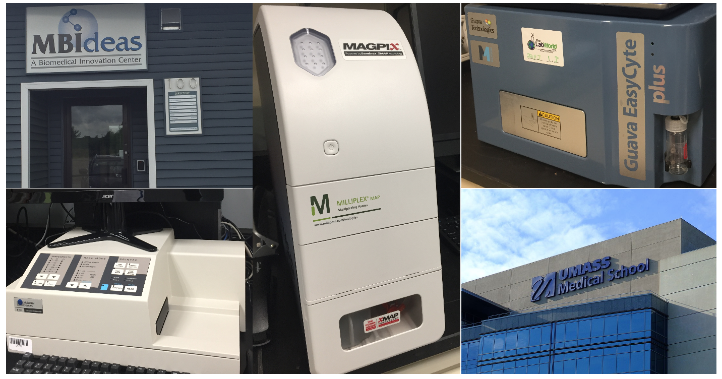
We have in house instrumentation for multiplexed soluble biomarkers (ELISA, Luminex), as well as cell-surface biomarkers (multiwell plate flow cytometry). We also have access to advanced microscopy and other instrumentation at the University of Massachusetts Core Research Facilities (CRFs) at their Worcester, Lowell and Amherst campuses.